Multimodal Generative AI in Radiology: Automating Reports and Annotations
- Mark Chomiczewski
- 26 July 2026
- 0 Comments
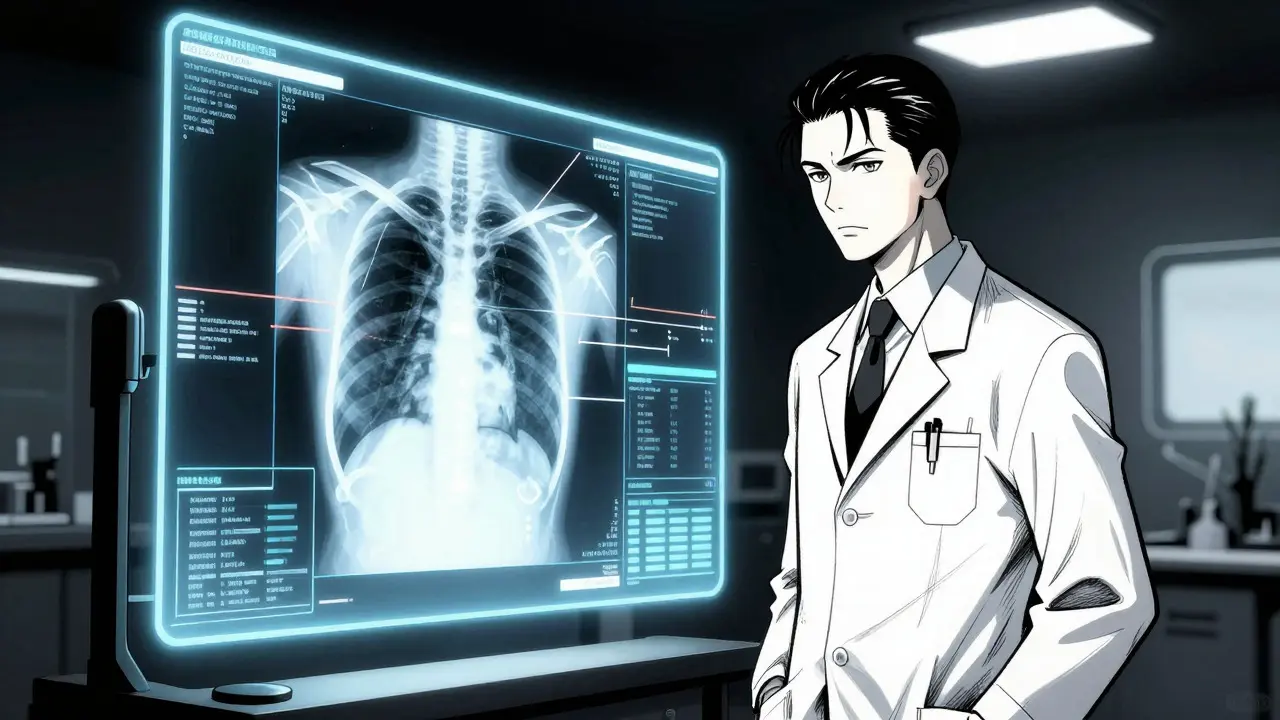
Imagine a world where your chest X-ray is analyzed, annotated, and turned into a detailed clinical report in seconds, not minutes. This isn't science fiction anymore; it's the current reality of Multimodal Generative AI, specifically applied to healthcare imaging through systems known as Generative Medical Image Interpretation (GenMI). As of late 2025, this technology has moved from academic curiosity to active pilot programs in nearly 80% of major US academic radiology departments. But does it actually work, or is it just another tech hype cycle? The answer lies in how these models process visual data alongside clinical context to assist-rather than replace-the human radiologist.
How Multimodal Generative AI Works in Radiology
To understand why GenMI is such a big deal, you need to look under the hood. Traditional AI in medicine often looked at images in isolation. A model might detect a nodule on a lung scan but ignore the patient’s history of smoking or recent trauma. Generative Medical Image Interpretation (GenMI) changes this by using transformer-based architectures that synthesize visual medical data with rich clinical context.
These systems typically use a dual-pathway approach. On one side, a Vision Transformer (ViT) processes the medical image-whether it’s an X-ray, CT, or MRI-at high resolution (usually 512x512 pixels). On the other side, a text encoder ingests clinical metadata, patient history, and procedural notes. The magic happens when these two streams merge. The model doesn’t just see pixels; it understands the narrative. For example, if a patient has a history of cancer, the AI pays closer attention to subtle changes in tissue density that might indicate recurrence. Microsoft’s CXRReportGen, for instance, uses 12 transformer layers with 16 attention heads to achieve 94.7% accuracy in identifying critical findings on chest X-rays, compared to 96.2% for human radiologists in internal validations.
But what about 3D scans like CTs or MRIs? Processing hundreds of slices individually is inefficient. Researchers at Stanford and Harvard proposed treating 3D medical images as video sequences. By converting a 300-slice CT study into a 20-frame "video," existing video-language models can analyze temporal and spatial relationships more effectively. This approach has shown an 89.3% accuracy rate in 3D interpretation, significantly outperforming standard vision-language models which hover around 82.7%. However, this method demands serious computing power, requiring NVIDIA A100 GPUs with 80GB VRAM and substantial system RAM.
The Real-World Impact: Speed vs. Accuracy
The primary driver for adopting GenMI is simple: there aren’t enough radiologists. The World Health Organization estimates a global shortage of 250,000 specialists by 2030. Hospitals are drowning in uninterpreted scans. GenMI offers a lifeline by drastically reducing documentation time. A prospective cohort study published in JAMA Network Open in 2024 found that these systems cut average report generation time from 14.7 minutes to just 8.2 minutes per study. That’s a 45% reduction in workload for routine cases.
But speed means nothing if accuracy suffers. Here, the picture gets nuanced. For uncomplicated cases-like a straightforward pneumonia check or a broken bone-GenMI systems shine, achieving up to 92.4% clinical accuracy. They excel at generating structured reports that highlight obvious pathologies. However, complexity drops performance. When dealing with oncology staging that requires correlating PET, CT, and MRI data, accuracy falls to 76.3%. More concerning is the issue of "nuanced impressions." In 31.7% of cases, current models fail to capture subtle details, such as the progression of a small pneumothorax or the texture of a benign vs. malignant nodule. Dr. Curtis Langlotz of Stanford University warned that these "dangerous blind spots" remain a significant hurdle for full autonomous deployment.
| Metric | Human Radiologist | GenMI System (e.g., CXRReportGen) |
|---|---|---|
| Average Report Time | 14.7 minutes | 8.2 minutes |
| Critical Finding Accuracy (Chest X-Ray) | 96.2% | 94.7% |
| Complex Multi-Modal Accuracy | High (Contextual Integration) | 76.3% (Struggles with Correlation) |
| Nuanced Impression Generation | Excellent | Poor (31.7% failure rate) |
| Fatigue Impact | High (Night shifts, volume) | None (Consistent performance) |
Key Players and Architectural Approaches
The market for medical GenMI is heating up fast, valued at $2.47 billion in Q3 2025 and projected to nearly quadruple by 2028. Three giants dominate the landscape: Microsoft, NVIDIA, and Google Health AI, controlling roughly 69% of the market. But their approaches differ significantly.
Microsoft leads with its Dragon Copilot and Foundry healthcare models. Their CXRReportGen Premium is the gold standard for chest X-rays, offering improved tuberculosis detection (93.2% accuracy). It’s expensive-$185,000 annually per institution-but it comes with robust documentation and integration support. On the other hand, open-source alternatives like RadFM from Stanford offer a cheaper entry point at $42,000, though they lag behind in critical finding detection (83.5% vs. 94.7%).
NVIDIA focuses on infrastructure with its CLARA platform. Their recent release of CLARA GenMI 2.0 emphasizes real-time procedural guidance, helping technicians position patients correctly during scans to reduce repeat rates. Meanwhile, startups are experimenting with Graph Neural Networks (GNNs) to map anatomical relationships more precisely, though these are less common in commercial deployments.
Choosing between these options depends on your hospital’s size and budget. Large academic centers with deep pockets often opt for Microsoft’s comprehensive suite, while smaller clinics might start with cloud-based solutions or open-source models to test the waters. However, remember that implementation isn’t plug-and-play. Integrating these tools with existing Electronic Health Records (EHRs) via HL7/FHIR standards can take over two months and require dedicated IT staff.
Challenges: Bias, Black Boxes, and Liability
If GenMI is so great, why isn’t everyone using it yet? The barriers are significant. First, there’s the "black box" problem. 76.4% of proprietary models lack transparency, meaning doctors don’t know *why* the AI made a specific recommendation. In medicine, explainability is crucial for trust and legal defensibility.
Second, bias remains a critical flaw. Training datasets for most GenMI models represent only 32.7% of global population diversity. This means the AI might perform well on Caucasian male patients but struggle with different ethnicities, ages, or body types. The FDA recognized this risk in August 2025, issuing new guidelines (FDA Guidance #2025-087) that require 95% sensitivity for critical findings and prospective validation across 10,000+ diverse studies before approval.
Liability is another headache. If the AI misses a tumor, who is responsible? The doctor who signed off on the report? The hospital that bought the software? Or the vendor who trained the model? Currently, 78% of malpractice insurers require explicit disclaimers stating that GenMI is an assistive tool, not a diagnostic authority. This reinforces the "human-in-the-loop" paradigm, where the AI drafts the report, but the radiologist must review and approve it. While this reduces error rates from 4.7% to 0.9%, it also limits the potential time savings.
Implementation Roadmap for Healthcare Providers
For hospitals considering GenMI adoption, the journey is complex. Based on the RSNA 2025 Implementation Survey, successful deployments take an average of 6.2 months. Here’s what you need to prepare for:
- Infrastructure Audit: Ensure you have the necessary GPU resources or cloud partnerships. Local processing requires NVIDIA A100s or similar high-end hardware.
- DICOM and EHR Integration: Plan for 3-5 full-time equivalent (FTE) engineers to handle data pipelines. Interoperability with systems like Epic or Cerner is non-negotiable.
- Clinician Training: Radiologists need 8-12 weeks of supervised use to become proficient. Focus on teaching them how to spot AI errors, particularly in nuanced impressions.
- Pilot Program: Start with low-risk, high-volume studies like chest X-rays or musculoskeletal exams. Avoid starting with complex oncology or neurology cases.
- Validation Framework: Establish internal metrics to track AI performance against human benchmarks. Regularly audit false positives and negatives.
User feedback suggests that early adopters see immediate benefits in reduced documentation fatigue. 84.7% of radiologists in the JAMA study reported feeling less burned out after implementing GenMI. However, 61.2% expressed concerns about "alert fatigue" from excessive false positives, which can desensitize clinicians to real warnings. Balancing sensitivity and specificity is key to maintaining trust.
The Future of Radiology: Assistive, Not Autonomous
Where is this technology heading? The consensus among experts is cautious optimism. GenMI will likely never fully replace radiologists, especially for complex cases requiring holistic judgment. Instead, it will become an indispensable co-pilot. By 2027, 73.4% of systems are expected to integrate directly with EHRs, pulling in lab results and genetic data to provide even richer context. Patient-facing interactive reports, which link findings directly to image regions, are already in beta testing with high satisfaction rates.
As we move toward 2026 and beyond, the focus will shift from raw accuracy to workflow harmony. The goal isn’t just faster reports; it’s better care. By freeing radiologists from repetitive documentation, GenMI allows them to spend more time on complex diagnostics and patient communication. The technology is imperfect, biased, and legally ambiguous, but it’s here. And for hospitals struggling with staffing shortages, it’s not just an option-it’s a necessity.
What is Multimodal Generative AI in healthcare?
Multimodal Generative AI in healthcare refers to artificial intelligence systems that combine multiple types of data-such as medical images (X-rays, CTs), clinical notes, and patient history-to generate comprehensive outputs like radiology reports. Unlike traditional AI that looks at data in silos, multimodal AI synthesizes visual and textual information to mimic human diagnostic reasoning, improving both speed and contextual accuracy.
How accurate are GenMI systems compared to human radiologists?
For routine tasks like chest X-ray analysis, GenMI systems achieve approximately 94.7% accuracy in critical finding identification, closely matching human radiologists at 96.2%. However, accuracy drops to 76.3% for complex multi-modal cases requiring correlation across different imaging types. Additionally, these systems struggle with nuanced impressions, failing in about 31.7% of cases where subtle clinical judgment is required.
What are the main limitations of current GenMI technology?
The primary limitations include dataset bias, with training data representing only 32.7% of global population diversity, leading to potential inaccuracies in underrepresented groups. Other issues include "black box" opacity (76.4% of models lack transparency), difficulty in correlating longitudinal findings across multiple studies (22.4% failure rate), and challenges in generating nuanced clinical impressions for rare diseases.
How long does it take to implement GenMI in a hospital?
Successful deployment typically takes around 6.2 months on average. This timeline includes infrastructure setup, DICOM and EHR integration (which can add 2-3 months), and clinician training. Radiologists generally require 8-12 weeks of supervised use to reach proficiency, ensuring they can effectively review and correct AI-generated drafts.
Who are the leading providers of GenMI solutions?
The market is dominated by Microsoft (with Dragon Copilot and CXRReportGen), NVIDIA (CLARA platform), and Google Health AI, which collectively hold about 69% of the share. Microsoft’s CXRReportGen Premium is noted for high accuracy but comes at a higher cost ($185,000/year), while open-source alternatives like Stanford’s RadFM offer a more affordable entry point ($42,000/year) with slightly lower performance metrics.